General tips
- Erythema annulare centrifugium (EAC) is a chronic condition that usually presents as a reaction to an antigen.
- Many underlying conditions have been reported to be associated with EAC but in most of cases, the underlying antigen can`t be identified.
- It can occur at any age.

History taking:
History of infection
- Fungal infection
- Dermatophytoses, Candida, Penicillium in blue cheese.
- Bacterial infection
- Mycobacteria, streptococci, Escherichia coli, syphilis, Pseudomonas septicaemia.
- Viral infection
- Epstein–Barr virus, HIV, herpes simplex, herpes zoster , molluscum contagiosum, viral hepatitis.
- Parasitic infection
- Ascaris lumbricoides, Phthirus pubis.
History of drug taking
- Non‐steroidal anti‐inflammatory drugs.
- Antimalarials.
- Diuretics : hydrochlorothiazide, spironolactone.
- Antibiotics : Ampicillin, co‐trimoxazole, penicillins.
- Others : Acetazolomide, amitriptyline,cimetidine,finasteride, rituximab, pegylated interferon-α-2a plus ribavirin, ustekinumab.
History of malignancy (paraneoplastic erythema annulare centrifugum)
It typically precedes the clinical diagnosis of malignancy, and may recur with subsequent relapses.
- Skin tumor : Squamous cell carcinoma
- Hematological : Acute myelogenous leukaemia, chronic lymphocytic leukaemia , hypereosinophilic syndrome , lymphomas, myeloma.
- Internal : Breast, bronchial, naso‐pharyngeal, ovarian, prostatic and gastric carcinomas.
History of autoimmune diseases
- Autoimmune hepatitis, lupus erythematosus, polyglandular autoimmune disease type 1 , relapsing polychondritis , Sjögren syndrome.
Adult female patient
- Pregnancy
- Autoimmune progesterone dermatitis.
Others
- Appendicitis , cholestatic liver disease, inflammatory bowel disease, sarcoidosis, hyperthyoidism.
Examination findings
Two type of erythema annulare centrifugium have been descibed
- Superficial type:
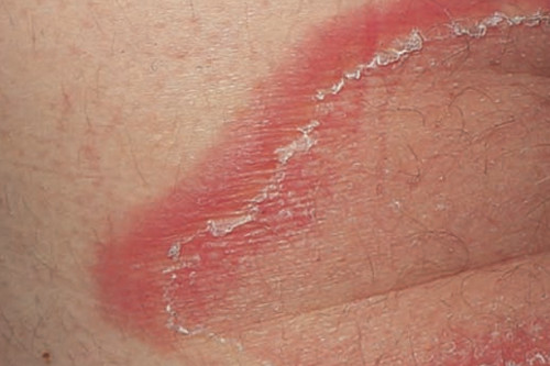
- It usually starts as firm pink papules that expand centrifugally followed by central clearing.
- It enlarges gradually and it may reach few centrimetres in diameter.
- It may form annular, polycyclic palques or patches or incomplete arcs.
- The inner margin shows trailing scale (desquamation).
- The lesions may be pruritic.
- Vesicles may be sometimes seen on the border.
- Deep type:
- The edge is slightly elevated ane the lesions are more indurated.
- No trailin scales.
- No pruritus.
Affected body parts
- The trunk and lower extremities are most commonly affected.
Investigations
- Exclude dermatophytes infection because it is one of the most common associations( KOH- culture)
- Biopsy with direct immunofluorescence to exclude other similar annular diseases.
- Full blood count : to evaluate a suspected underlying infection (neutrophilia with bacterial infection; eosinophilia with parasitic infection or hypereosinophilic syndrome).
- Liver and thyroid function tests : to exclude hepatitis and hyperthyoidism.
- Chest X‐ray : to exclude tuberculosis, malignancy (primary or metastatic), sarcoidosis, or lymphoma.
- An antinuclear antibody profile for lupus.
- Serum angiotensin‐converting enzyme (ACE): for sarcoidosis.
- HIV serology
- If malignancy suspected, PET-CT should be considered.
Erythema annulare centrifugium should be differentiatied from the different annular conditions such as:
- Tinea corporis and tinea imbricata
- Clinically,pruritic and scaly. The scales are usually on the leading edge not the trailing one.
- KOH and culture can exclude fungal infection.
- Granuloma annulare
- No scales.
- Biopsy can be done to differentiate, granuloma annulare shows an interstitial and/or palisaded, histiocytic infiltrate with or without collagen necrobiosis.
- Erythema marginatum
- The patient usually will be a child with acute rheumatic fever.
- Annular erythematous plaques appear rapidly then fade over the course of hours to several days.
- Erythema migrans
- Expanding erythematous patch usually surrounding a central bite site with central clearing, occurs ~1-2 weeks after tick bite.
- It disappears typically within 4 weeks without treatment.
- Patients may have associated fatigue and lymphadenopathy.
- Histopathology and serology can be used to differntiate.
- Erythema gyratum repens
- Gyrate polycyclic plaques with trailing scale.
- Itchy.
- A strong association with Various malignancies, bronchogenic carcinoma is the most common.
- Pathologically different from EAC.
- Erythema papulatum centrifugum
- Itchy and sweat related condition.
- It presents as annular plaues with the border formed of multiple small papules.
- Pathologically,biopsy shows inflammation around dermal and intraepidermal eccrine ducts.
- Erythema multiforme
- acute presentation of target lesions affecting mostly the acral parts.
- Mucous memebranes may be affected.
- Lesions may show vesicles or bullae.
- Subacute cutaneous lupus erythematosus
- The lesions usually affects the sun exposed skin.
- Biopsy and serology can be done to differentiate.
- Annular secondary syphilis
- If suspected, rapid plasma reagin (RPR) can be done.
- Annular sarcoidosis
- Naked granuloma can be seen on biopsy.
- Other organ affection like pulmonary sarcoidosis may be present.
- Necrolytic migratory erythema
- It is assocaited with Glucagonoma syndrome, pancreatic tumor can be detected by CT scan.
- Patients may be present with weight loss, diarrhea, anemia and diabetes mellitus.
- The distribution of the lesions tends to be perioral, perianal, and in the inguinal folds
- Annular urticaria
- Urticarial lesions usually fades in less than 24 hours.
- Urticarial phase of bullous pemphigoid
- Older patients are affected.
- Biopsy can be used to confirm the diagnosis.
Course and prognosis
- Lesions of erythema annulare centrifugum`s duration is variable.
- It may last form days to decades.
How to diagnose erythema annulare centrifugum?
- Erythematous annular lesions expanding centrifugally followed by clearing of the center.
- Trailing scales on the inner edge of the lesion border.
- Pathological features of superficial type is not specific, but deep type may show tight aggregate around vessels, the so-called “coat sleeve” appearance.
- Histopathogical features can help to exclude other annular diseases.