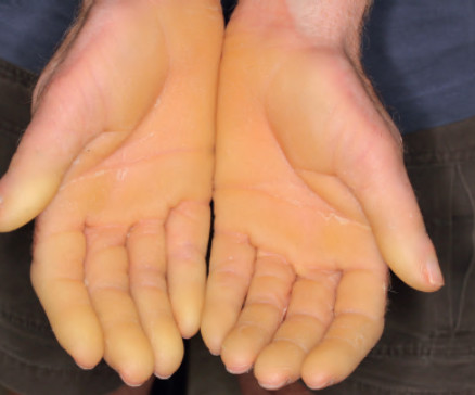
Yellow to orange skin lesions in dermatology
Endogenous or exogenous pigment
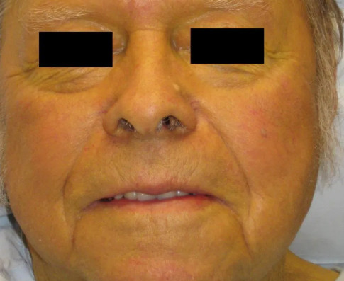
- Hyperbilirubinemia(Jaundice)
- Ecchymoses.
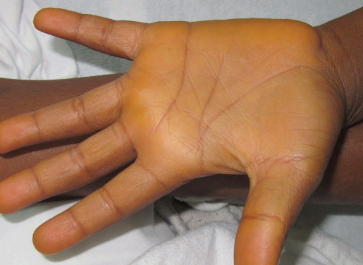
- Carotenemia.
- Drug induced pigmentation (e.g. quinacrine).
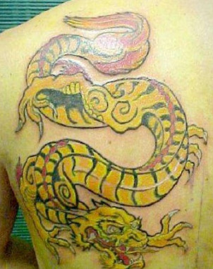
- Cadmium tattoo.
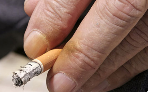
- Tobacco staining.
- Yellow chromonychia.
Skin disorders of elastic tissue
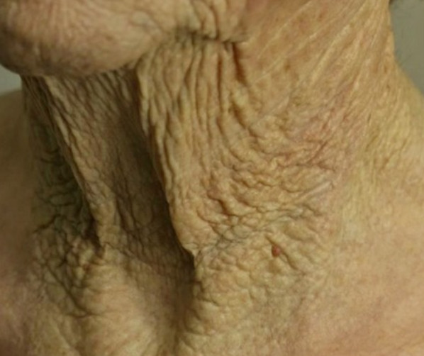
- Solar elastosis.
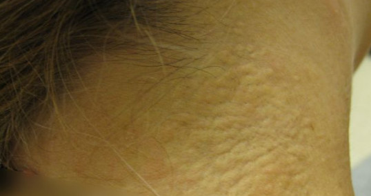
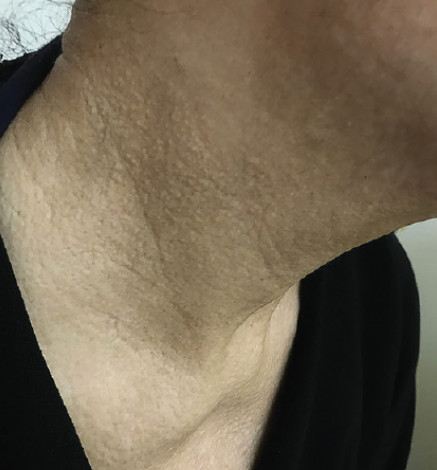
- Cutis rhomboidalis nuchae.
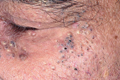
- Favre–Racouchot syndrome.
- Pseudoxanthoma elasticum.
- Papillary dermal elastolysis.
- Mid-dermal elastolysis.
- Keratoelastoidosis marginalis.
- Connective tissue naevus.
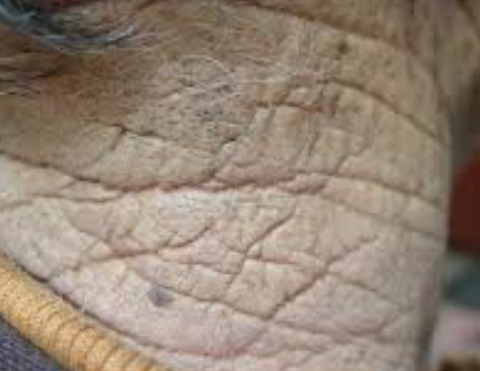
Cutis rhomboidalis nuchae.
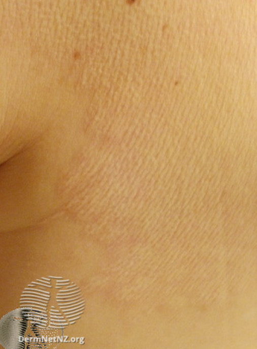
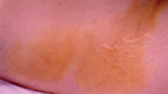
Connective tissue nevus
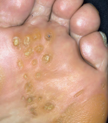
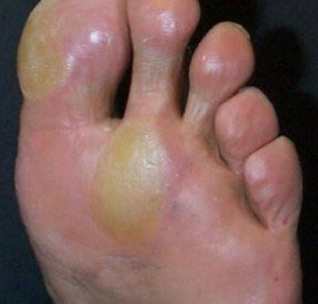
Skin disorders due to keratin accumulation
- Calluses.
- Corns.
- Keratoderma.
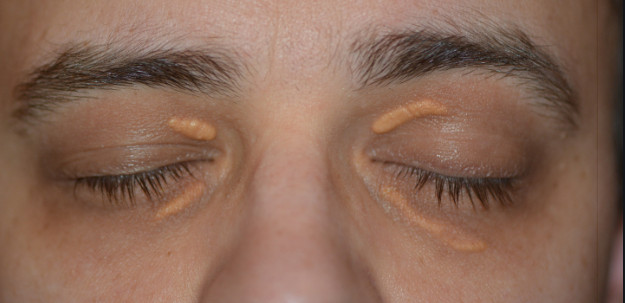
Skin disorders due to lipid accumulation(Xanthomatosis)
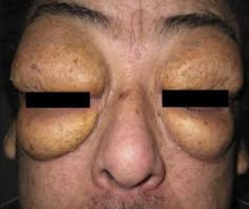
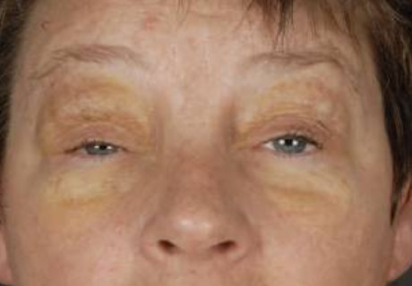
- Xanthelasma.
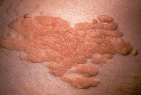
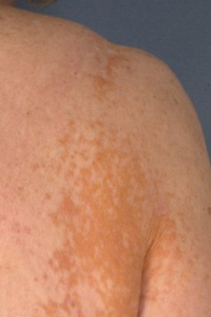
- Plane xanthoma.
- Eruptive xanthoma.
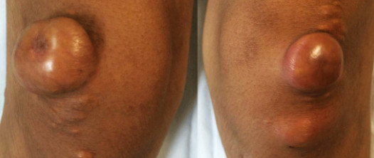
- Tuberous xanthoma.
Cutaneous histiocytosis
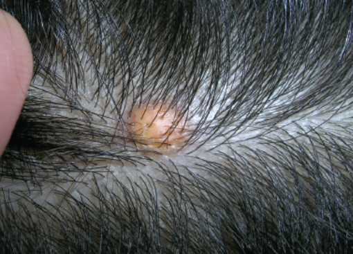
- Juvenile xanthogranuloma.
- Solitary reticulohistiocytoma.
- Benign cephalic histiocytosis.
- Langerhans cell histiocytosis.
- Necrobiotic xanthogranuloma.
- Xanthoma Disseminatum.
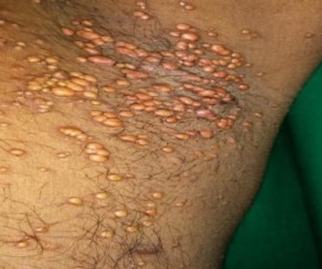
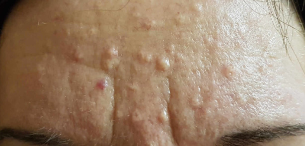
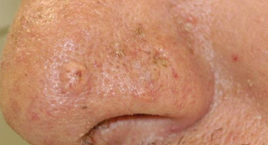
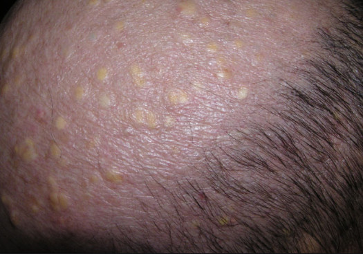
Sebocytes related conditions
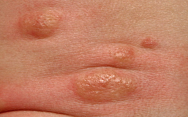
- Sebaceous hyperplasia.
- Sebaceous adenoma.
- Sebaceoma.
- Sebaceous carcinoma.
- Steatocystoma multiplex.
- Naevus sebaceous of Jadassohn.
Infections
- Crusted impetigo
- Onychomycosis
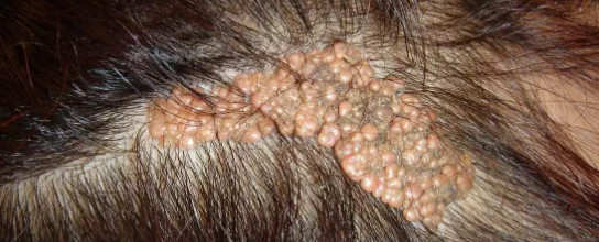
- Trichomycosis axillaris and pubis.
Others
- Gouty tophi.
- Verruciform xanthoma.
- Lipoid proteinosis.
- Mastocytoma
- Goltz syndrome.
- Lichen aureus.
- Colloid milium.
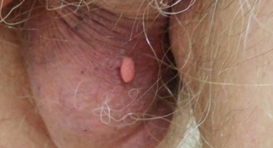
- Piezogenic pedal papules
- Yellow nail syndrome.
- Extranodal marginal zone B-cell lymphoma.
References
- Frew, J.W., Murrell, D.F. and Haber, R.M. (2015), Fifty shades of yellow: a review of the xanthodermatoses. Int J Dermatol, 54: 1109-1123. doi:10.1111/ijd.12945.
- Bañuls, J., Arribas, P., Berbegal, L., DeLeón, F. J., Francés, L., & Zaballos, P. (2015). Yellow and orange in cutaneous lesions: clinical and dermoscopic data. Journal of the European Academy of Dermatology and Venereology, 29(12), 2317-2325.
- https://dermnetnz.org/
#Yellowish skin lesions in dermatology #Yellowish skin lesions.